Big science on a big scale; Microscope brings new power to cancer fight
Published 10:54 am Wednesday, June 1, 2016
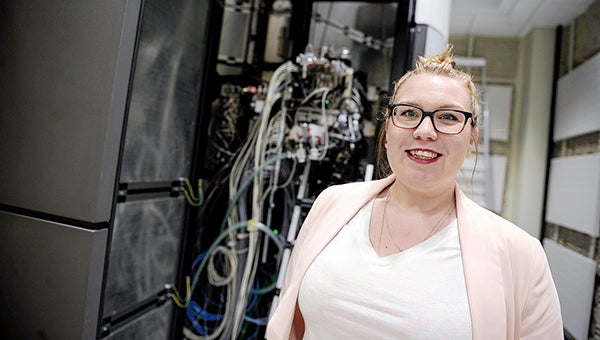
Dr. Anna Sundborger, assistant professor and section leader for the cryo-electron microscope, stands in front of the hulking machine, now being installed in The Institute. — Photos by Eric Johnson/photodesk@austindailyherald.com
Look out cancer cells, there’s a new microscope in town.
As The Hormel Institute celebrates the grand opening of its $27 million expansion Wednesday, it’s also celebrating new equipment that will benefit scientists at The Institute and around the region.
The Institute recently purchased a $4.3 million cryo-electron microscope that will help scientists analyze the structure of proteins and amino acids.
Assistant professor and section leader for the cryo-electron microscope Dr. Anna Sundborger said the microscope will help determine how a protein is structured, how different proteins bind to each other and eventually how to design drugs that can fight cancer cells.
“Electro-microscopy has been around for a long time as kind of a way to look at things inside a cell at really high magnification,” Sundborger said. “… You can see, basically the different compartments of the cell and you can look at how cells interact with each other, you can look at how bacteria and viruses interact with cells.”
Previous cameras only interpreted the electrons that were passing through the sample, but now the camera is counting the number of electrons which allows for more detail in the pictures, Sundborger said.
Sensitive instrumentation
The microscope was delivered in late March in 80 crates that filled an entire hallway outside the microscope room. It takes three months to put together, and workers from Netherlands-based FEI are in Austin assembling it.
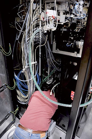
A technician works on assembling the $4.3 million cryo-electron microscope at the The Hormel Institute.
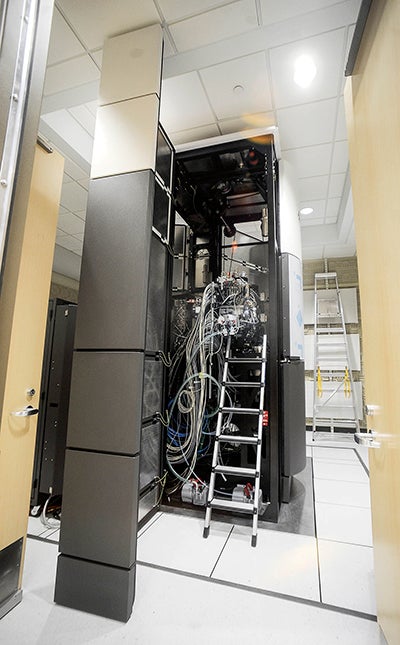
In order for the camera to capture images properly, the microscope is being installed in a specialized room so the signals and tests won’t be interrupted. The room that houses the cryo-electron microscope was specially built to control sound, vibration and temperature.
“These microscopes are extremely sensitive. They’re sensitive to everything, to humidity, to temperature,” Sundborger said. “They have to have a perfectly controlled environment.”
A special pit was built below the room housing the microscope and measures were taken to ensure a constant temperature for 24 hours, which wasn’t necessarily easy, according to Building Coordinator Craig Jones.
“That’s quite difficult for your standard HVAC system,” he said.
Sundborger added the signal to noise ratio is important and very little is done to the samples to give them contrast in the image. The images are produced in grayscale and scientists must be able to distinguish it from the background, but the protein will pop out as a darker structure in that gray area.
“Anything you do in that room to cause interference with the camera is making it harder for the computer to distinguish, so it just brings in more noise,” Sundborger said. “So if you have music playing, your camera is going to register that and it’s going to make your image noisier, so it’s going to be harder for the computer to distinguish what kind of sample you have.”
Jones noted no one will be in the room with the microscope when tests are being performed.
How it works
The microscope is able to take a number of samples at one time, which are frozen in liquid nitrogen, hence the “cryo” part. The microscope can analyze the samples continuously for up to seven days on its own.
“You don’t even have to be in the room,” Sundborger said. “So you’re sitting in the room behind it and running the microscope, but I could be sitting here or at my house controlling the microscope, so that’s pretty cool.”
According to Associate Director Ann Bode, it’s important for samples to be prepared in sub-zero conditions because it allows scientists to visualize the cell and protein structure through crystallography. Many proteins can be crystalized through various processes, but those alter the sample’s natural structure and can degrade it over time. But the samples won’t need to be altered when using the cryo-electron microscope.
“The cryo-electron microscope allows us to use the native protein,” Bode said. “We don’t have to alter it all, so then you can visualize … the natural structure, the formation that occurs in nature rather than trying to manipulate it into something that isn’t real.”
Sundborger added that cryogenic technology has been revolutionized in the past five years. Instead of crystallizing samples or making them black and white, the sample is fixated in ice, or liquid nitrogen, which keeps everything intact instead of destroying it.
“You’re taking the natural sample and you’re just freezing them really quickly, instead of fixating them in different things that basically destroy everything, but keeps it intact,” Sundborger said.
The camera, which allows visualization at the atomic level, will take tens of thousands of pictures of proteins’ structures, compare and average the pictures and then using a special software, create 3D models to see how the protein is organized.
The ultimate goal is to see how proteins bind to each other and how drugs bind to the proteins.
The $4.3 million cryo-electron microscope at The Hormel Institute, when finished, will allow scientists to examine protein structure at the very smallest of levels.
“An example is that you have a cell and then on the surface of the cell has all these proteins and they’re called cell-surface receptors and in the context of cancer, for instance, you can have these drugs that bind to these receptors,” Sundborger said.
The designed drugs can also block or promote a function within the cell, which will be negative for the cancer cell, she added.
“It’s essentially going to kill the cancer cell, but to be able to assign the perfect drug, you need to know exactly what that protein receptor looks like,” Sundborger said. “So you need to have as good as a protein structure as possible.”
For now, Sundborger plans to use the microscope to see how cells react in a normal environment and analyze proteins that are involved in different cancer pathways. She said this is as equally important as studying cancer, because the pathways are involved in cancer.
“What could have happened is that you had a bacterial infection and it messed up some little protein pathway and therefore, 20 years later, you had cancer, because that little pathway led to some other pathway that was disrupted and so on and so on,” Sundborger said. “Cancer is a really complicated disease when it comes to how it starts, but the end result is always the same, that you have this uncontrolled cell growth.”
Shared use
Sundborger estimated there were possibly about five to ten cryo-electron microscopes in use right now, but they are still pretty rare, and Bode agreed the microscope and its biological use is unique to the area.
“This particular instrument is the only one for biological applications in this whole region,” Bode said. “In any state around us, there’s none other like it.”
A cryo-electron microscope is being used in Minneapolis for material science.
“It’s a huge deal if you have one of them,” Sundborger said. “So we’re going to be very unique. I don’t think they even have one in Chicago. Not at Mayo, they don’t have one. It’s going to be a lot of people coming here probably to use it.”
The microscope won’t just be used by Institute scientists. Scientists and researchers from Institute partners like the University of Minnesota and Mayo Clinic plan to use the microscope, and The Institute has already had inquiries from scientists in Minnesota, Iowa, and the South Dakota asking if they can buy time for their own studies.
“This is going to be something that’s really quite phenomenal for the Institute,” Bode said.
That shared use is part of what allowed The Institute to purchase the expensive equipment.
Sundborger has worked at The Hormel Institute for the past three months and plans to use her knowledge of the microscope to conduct research. She has a Ph.D. in neuroscience and a masters degree in medical biology. She is originally from Stockholm, Sweden.
While Sundborger was hired to lead a research team utilizing the new technology, more support staff is planned to help manage the specialized equipment.
“We’re doing a search now for more faculty and staff,” Bode said.
—Jason Schoonover contributed to this report.